


SARS-CoV-2 Nucleocapsid Proteins
| Product | Unit size | Cat. code | Docs. | Price | |
|---|---|---|---|---|---|
|
Nucleocapsid-His SARS-CoV-2 Nucleocapsid-His fusion protein |
Show product |
50 µg |
his-sars2-n
|
||
|
Nucleocapsid-Fc SARS-CoV-2 Nucleocapsid-Fc fusion protein |
Show product |
50 µg |
fc-sars2-n
|
SARS-CoV-2 Nucleocapsid with C-term His- or Fc-tag
Protein description

Potential applications of soluble tagged Nucleocapsid proteins
 InvivoGen also offers:
• Soluble Spike S1 proteins
• Soluble Spike RBD proteins
• Soluble Human ACE2 protein
The SARS-CoV-2 (2019-nCoV) Nucleocapsid (N) is a structural protein that plays important roles in the viral life cycle including replication, transcription, and genome packaging [1]. The SARS-CoV-2 N features two important NTD and CTD functional domains [1-6]. NTD interacts with both the RNA genome and Membrane/Matrix (M) proteins to form virion particles. The N protein interaction with the RNA forms the virus ribonucleoprotein core which is packed as a helical “beads-on-a-string” conformation. CTD allows RNA synthesis through binding of the replication-transcription complexes (RTCs), oligomerization of multiple N proteins through its dimerization domain, and genome incorporation into the new virion.
N is a major immunogen of SARS-CoV-2. Indeed, elevated Anti-SARS-CoV-2 N IgG and IgM antibody titers have been reported in COVID-19 patients’ sera [7-9]. These observations make SARS-CoV-2 N an attractive tool for early diagnosis [7-9], and treatment strategies [3].
Nucleocapsid-His and Nucleocapsid-Fc were generated by fusing the full-length SARS-CoV-2 N [M1-A419] to a C-terminal poly-histidine sequence and human IgG1 Fc region, respectively. Of note, the SARS-CoV-2 viral sequence used is from the Wuhan-Hu-1 (D614) isolate.
Nucleocapsid-His and Nucleocapsid-Fc have been produced in CHO cells and HEK293 cells, respectively, and have been purified by affinity chromatography (See Specifications for more information).
Applications
- Vaccination studies: using combinations of Nucleocapsid protein antigens and adjuvants
- Antibody screening: finding anti-Nucleocapsid antibodies in COVID-19 patients' sera
- Inhibitor screening: finding small molecules able to block the SARS-CoV-2 Nucleocapsid interaction with replication-transcription complexes (RTCs)
Quality control
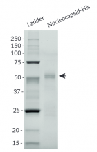
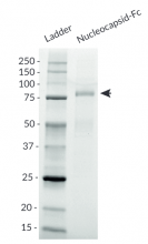
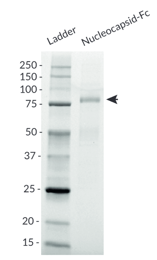
- Size and purity confirmed by SDS-PAGE
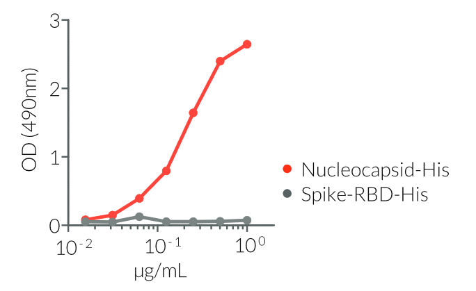
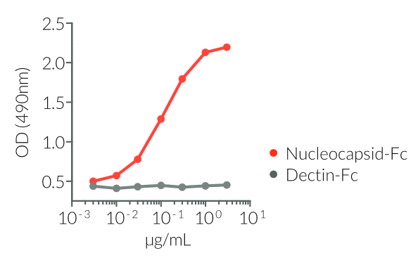
- Protein validated by ELISA using a coated anti-SARS Nucleocapsid antibody
![]() Learn more about SARS-CoV-2 infection cycle, immune responses, and potential therapeutics.
Learn more about SARS-CoV-2 infection cycle, immune responses, and potential therapeutics.
References
1. Mu, J. et al., 2020. SARS-CoV-2-encoded nucleocapsid protein acts as a viral suppressor of RNA interference in cells. Sci China Life Sci 63, 1-4.
2. Chang C. et al., 2006. Modular organization of SARS coronavirus nucleocapsid protein. J. Biom. Sci. 13:59-72.
3. Krokhin O. et al., 2003. Mass spectrometric characterization of proteins from the SARS virus. Mol. & Cell. Prot. 2:346-356.
4. Cubuk, J. et al., 2020. The SARS-CoV-2 nucleocapsid protein is dynamic, disordered, and phase separates with RNA. bioRxiv. doi:10.1101/2020.06.17.158121.
5. Kang, S.et al., 2020. Crystal structure of SARS-CoV-2 nucleocapsid protein RNA binding domain reveals potential unique drug targeting sites. Acta Pharm Sin B. doi:10.1016/j.apsb.2020.04.009.
6. Khan, M.T. et al., 2020. SARS-CoV-2 nucleocapsid and Nsp3 binding: an in silico study. Arch Microbiol. doi: 10.1007/s00203-020-01998-6.
7. Liu, W. et al., 2020. Evaluation of Nucleocapsid and Spike Protein-Based Enzyme-Linked Immunosorbent Assays for Detecting Antibodies against SARS-CoV-2. J Clin Microbiol 58.
8. Guo L. et al., 2020. Profiling Early Humoral Response to Diagnose Novel Coronavirus Disease (COVID-19). Clinical Infectious Diseases. 71(15) :778-785.
9. To K. K-W. et al., 2020. Temporal profiles of viral load in posterior oropharyngeal saliva samples and serum antibody responses during infection by SARS-CoV-2: an observational cohort study. The Lancet Infectious Diseases. 20(5):565-574.
Figures


Specifications
Nucleocapsid-His
- Protein construction: Full-length Nuceocapsid with a C-terminal poly-histidine tag
- Accession sequence: P0DTC9
- Species: SARS-CoV-2 (2019-nCoV); Wuhan-Hu-1 (D614) isolate
- Tag: C-terminal poly-histidine (6 x His)
- Total protein size: 430 a.a. (secreted form)
- Molecular weight: ~ 52 kDa (SDS PAGE gel)
- Purification: Ni2+ affinity chromatography
- Purity: >95% (SDS PAGE)
-
Quality control:
- The protein has been validated by ELISA upon incubation with an Anti-SARS Nucleocapsid antibody.
- The absence of bacterial contamination (e.g. lipoproteins and endotoxins) has been confirmed using HEK-Blue™ TLR2 and HEK-Blue™ TLR4 cellular assays.
Nucleocapsid-Fc
- Protein construction: Full-length Nuceocapsid with a C-terminal human IgG1 Fc tag
- Accession sequence: P0DTC9
- Species: SARS-CoV-2 (2019-nCoV); Wuhan-Hu-1 (D614) isolate
- Tag: C-terminal human IgG1 Fc
- Total protein size: 669 a.a. (secreted form)
- Molecular weight: ~ 79 kDa (SDS PAGE)
- Purification: Protein A affinity chromatography
- Purity: >95% (SDS PAGE)
-
Quality control:
- The protein has been validated by ELISA upon incubation with an Anti-SARS Nucleocapsid antibody.
- The absence of bacterial contamination (e.g. lipoproteins and endotoxins) has been confirmed using HEK-Blue™ TLR2 and HEK-Blue™ TLR4 cellular assays.
Contents
Nucleocapsid-His and Nucleocapsid-Fc contents:
- 50 μg of lyophilized protein
- 1.5 ml of endotoxin-free water
![]() The product is shipped at room temperature.
The product is shipped at room temperature.
![]() Lyophilized protein should be stored at -20 ̊C.
Lyophilized protein should be stored at -20 ̊C.
![]() Resuspended protein is stable up to 1 month when stored at 4°C, and 1 year when stored at -20°C
Resuspended protein is stable up to 1 month when stored at 4°C, and 1 year when stored at -20°C
Avoid repeated freeze-thaw cycles.
Back to the top